Lindsay Holsen is theCEC/RTC summer intern, visiting us from Lawrence University in Appleton, WI. In addition to community engagement and research translation work, Lindsay is helping Duke doctoral student Tess Leuthner in Dr. Joel Meyer’s lab with a research project on soil nematodes and contaminated soil.
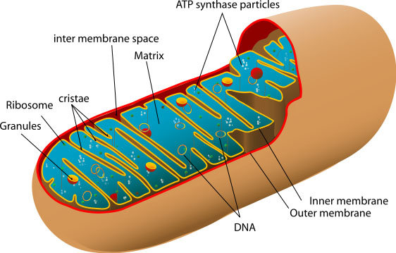
Figure 1. Mitochondria (Image: https://www.umdf.org/what-is-mitochondrial-disease/)
Scientists have theorized that one single, incredibly improbable event sparked a major change in the evolution of eukaryotic organisms.
The free-floating prokaryote precursor to the present-day mitochondria was engulfed by a eukaryote (a cell that contains a nucleus). Inside the eukaryote, the swallowed-up prokaryote was not destroyed; instead, the independent cells formed an unexpected mutual symbiosis that has developed into the sub-cellular organelles known as mitochondria within eukaryotic cells.
Thanks to the prokaryotic origins of mitochondria, they have their own genome inside a double membrane barrier. One of the primary roles of this unique organelle, aside from its role in cellular signaling, is its ability to synthesize ATP, a form of cellular energy that is available to the cell. This is a collaborative process of many proteins. The proteins along the membranes exchange and transport electrons, creating a proton gradient that converts of energy inputs into a useful form of cellular energy, ATP.
Advances in biological science, especially those within the past 30 years, have elucidated far greater complexity and function for the “power house of the cell,” a title introduced by Philip Siekevitz in 1957.
Part of escaping the singular functionality perception of mitochondria is recognizing that there can be thousands of uniquely shaped mitochondria in one cell. They form a dynamic network that is dependent upon the energy demands on the cell. Environmental conditions and cellular signaling can determine the rates of fusion, fission and mitophagy.
 Figure 2. Mitochondrial Dynamics
Image: http://www.acnr.co.uk/2014/04/mitochondrial-dynamics-as-a-potential-therapeutic-target-for-parkinsons-disease/
Mitochondrial fusion, fission, and mitophagy are responsive to environmental stressors. Those stressors can be part of the cellular process and alter cellular signaling from the mitochondria, but some stressors, known as “mitotoxicants” (toxicants affecting the mitochondria) have been shown to contribute to mitochondrial dysfunction and diseases, such as Parkinson’s, as a result of perturbations in mitochondrial dynamics. The chemicals do not actually cause the mutations that inhibit fission and fusion, but genetic modification of proteins causes deficiencies in these dynamic processes and increased sensitivity to certain chemicals (see Figure 2 of Luz’s paper) (Luz et al. 2017).
Many mitotoxicants are environmental toxicants, including certain Superfund chemicals. This emerging area of research is the basis for the Duke Superfund Research Center’s new Project 3, which addresses environmental mitotoxicant-induced mitochondrial dysfunction at critical developmental periods (reproductive age and early childhood), with the hypothesis that mitochondrial dysfunction resulting from these exposures will be transferable from parent to child. The project uses C.elegans as a model organism due to nematode’s quick growth to reproductive maturity (about 3 days), which allows for generational studies. Another post on the Superfund blog, Into the Lab, provides additional information about the benefits of using C.elegans as an in-vivo model.
Dr. Joel Meyer and Sherine Chan’s special issue of the journal Toxicology in June 2017, titled “Sources, mechanisms, and consequences of chemical-induced mitochondrial toxicity,” describes the emerging field of mitochondrial toxicity. As more diseases are being correlated, at least in part, to mitochondrial dysfunction induced by environmental contaminants, there is a glaring gap in the knowledge of the toxicological mechanisms that cause the diseases. Some other emerging concerns related to mitochondrial chemical-toxicity noted by Meyer and Chan are:
- Mitohormetics, which refers to an exposure to permanent or long-term diet and exercise habits and environmental chemicals among others that alter signalling and mitochondrial function that is beneficial at a low-dose, and harmful in high doses (Mattson, 2008)
- Environmental nonoxidative genotoxins that cause Mitochondrial DNA (mtDNA) mutations and disease
- Increased risk of mitotoxic exposure for those with pre-existing mitochondrial diseases or dysfunction
A recent publication by DUSRC Trainee Anthony Luz, “Deficiencies in mitochondrial dynamics sensitize Caenorhabditis elegans to arsenite and other mitochondrial toxicants by reducing mitochondrial adaptability” also used C. elegans to test 10 environmental toxicants, with arsenite as the primary concern, for their effects on genetic mutations and dysfunction in mitochondria (Luz et al. 2017). The study helped to define both the mechanistic complexity of determining and isolating the specific routes and causes of toxicity, especially in mitochondria. The paper complements Meyer and Chan’s concerns and elucidates the need for further mechanistic research, consideration of the effect of mitochondrial toxicants on previously compromised mitochondria, and impaired DNA damage removal in response to these types of toxins.
The human population faces a plethora of environmental exposures each day, with new, untested chemicals entering the market at a rapid pace. This makes mitochondrial toxicity an environmental health concern for further research in the health and toxicology fields as well as other disciplines that can work to address a wide variety of mitochondrial diseases and the mechanisms of mitochondrial toxicity.
References
Evans, A. (2016). Timeline: Mitochondria. Molecular Cell,61(5), 790. doi:10.1016/j.molcel.2016.02.015
Mattson, M. P. (2008). Hormesis Defined. Ageing Research Reviews, 7(1), 1–7. http://doi.org/10.1016/j.arr.2007.08.007
Meyer, J. N., & Chan, S. S. (2017). Sources, mechanisms, and consequences of chemical-induced mitochondrial toxicity.
Luz, A. L., Godebo, T. R., Smith, L. L., Leuthner, T. C., Maurer, L. L., & Meyer, J. N. (2017). Deficiencies in mitochondrial dynamics sensitize Caenorhabditis elegans to arsenite and other mitochondrial toxicants by reducing mitochondrial adaptability. Toxicology.
Youle, R. J., & Narendra, D. P. (2011). Mechanisms of mitophagy. Nature Reviews. Molecular Cell Biology, 12(1), 9–14. http://doi.org/10.1038/nrm3028